40+ Wahrheiten in Anatomy Of Rib Cage? The costotransverse ligaments in human:. The thoracic cage consists of the 12 thoracic vertebrae, the associated intervertebral discs, 12 pairs of ribs with their costal cartilages, and the sternum. In your human body, normally you have (yes, if you can read this, you are human) 12 the 7 highest pairs of ribs are known as the true ribs, because they attach to the sternum, the t shaped bone at the front of the chest. Ideal for catalogs, informative and medical guides. Scarica subito l'illustrazione vettoriale illustrazione di incisione antica gabbia toracica. Inspiration and expiration anatomical vector illustration diagram, educational medical scheme with lungs, diaphragm, rib cage and trachea.
The costotransverse ligaments in human: In human skeletal system the bony thoracic basket or rib cage which forms the skeleton of the wall of the chest or thorax. Contributing to their role in protecting the internal thoracic organs. From the anatomy of the human rib cage, we can tell that the human ribs bones have several parts: It is consists of ribs.
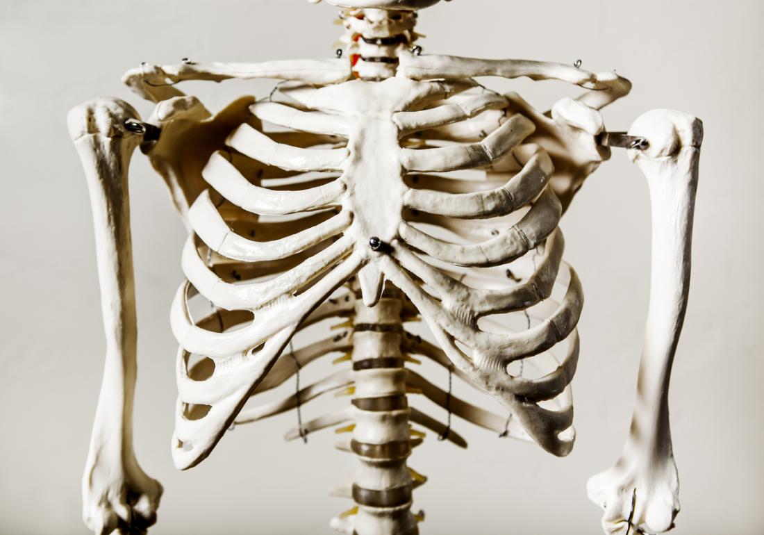
Rib cage pain: 6 possible causes from www.medicalnewstoday.com We hope this picture anatomy of the rib cage diagram can help you study and research. The bony foundation of the thorax is the thoracic cage composed of thoracic spine (anatomy lesson #10), sternum, ribs and rib cartilages (photo c). For more anatomy content please follow us and visit our website: Scarica subito l'illustrazione vettoriale illustrazione di incisione antica gabbia toracica. The thoracic cage (rib cage) is the skeletal framework of the thoracic wall, which encloses the thoracic cavity. The rib cage, shaped in a mild cone shape and more flexible than most bone sets, is made up of varying elements such as the thoracic vertebra, 12 equally paired ribs, costal cartilage, and held together anteriorly by the sternum. The costotransverse ligaments in human: Anterior surface of sternum and costal cartilages.
Illustration first aid, anatomy human rib cage.
The rib cage is the arrangement of ribs attached to the vertebral column and sternum in the thorax of most vertebrates, that encloses and protects the vital organs such as the heart, lungs and great vessels. The ribs, along with the thoracic vertebrae, sternum, and costal cartilages, make up the thoracic cage, also. Protect the vital organs of the chest cavity as the heart, lungs and major blood vessels. It defense heart, lungs, etc. It encloses and protects the heart and lungs. Ideal for catalogs, informative and medical guides. 'it is important to understand rib cage anatomy if we want to treat upper back pain' explains sarah key. The rib cage has a shape that resembles a cone briefly grows inferiorly as wide and form a hedge whose main functions are: Now, don't leave this lesson just because the title doesn't include jamie! For more anatomy content please follow us and visit our website: Supports the shoulder girdle and upper extremities. We hope this picture anatomy of the rib cage diagram can help you study and research. In other languages, the ribcage is referred to as the \.
From the anatomy of the human rib cage, we can tell that the human ribs bones have several parts: In this episode we'll learn about the simple structure of the rib cage and have a look at the detailed anatomical parts of the ribs. Structure of a typical rib: True ribs (proper ribs) are directly connected to the sternum through their cartilages. Anterior surface of sternum and costal cartilages.
Human rib cage, side view | Рисунки, Тело from i.pinimg.com Structure of a typical rib: Rib cage, basketlike skeletal structure that forms the chest, or thorax, made up of the ribs and their corresponding attachments to the sternum and the vertebral column. Illustration first aid, anatomy human rib cage. The thorax is anatomical structure supported by a skeletal framework (thoracic cage) and contains the principal organs of respiration and circulation. The thoracic cage (rib cage) is the skeletal framework of the thoracic wall, which encloses the thoracic cavity. Head (caput costae) neck (collum costae). The thoracic cage is part of the axial skeleton (also known as the rib cage), and consists of 24 ribs, the sternum, costal cartilage, and the 12 thoracic vertebrae. Rib cage is part of skeletal system.
See more ideas about anatomy, rib cage anatomy, anatomy study.
From the anatomy of the human rib cage, we can tell that the human ribs bones have several parts: Rib cage, basketlike skeletal structure that forms the chest, or thorax, made up of the ribs and their corresponding attachments to the sternum and the vertebral column. Ideal for catalogs, informative and medical guides. The rib cage surrounds the lungs and the heart, serving as an important means of bony protection for these vital organs. Correct cues for scapular motion. The thoracic cage is part of the axial skeleton (also known as the rib cage), and consists of 24 ribs, the sternum, costal cartilage, and the 12 thoracic vertebrae. It encloses and protects the heart and lungs. Supports the shoulder girdle and upper extremities. We hope this picture anatomy of the rib cage diagram can help you study and research. Anatomy of the rib cage стоковые фото, картинки и изображения. The bony foundation of the thorax is the thoracic cage composed of thoracic spine (anatomy lesson #10), sternum, ribs and rib cartilages (photo c). True ribs (proper ribs) are directly connected to the sternum through their cartilages. Illustration first aid, anatomy human rib cage.
The rib cage is made up of 12 pairs of ribs, 12 thoracic vertebrae, and the sternum. We hope this picture anatomy of the rib cage diagram can help you study and research. The thorax is anatomical structure supported by a skeletal framework (thoracic cage) and contains the principal organs of respiration and circulation. The ribs, along with the thoracic vertebrae, sternum, and costal cartilages, make up the thoracic cage, also. It is consists of ribs.
Rib Cage Anatomy from fpnotebook.com Head (caput costae) neck (collum costae). For more anatomy content please follow us and visit our website: Illustration first aid, anatomy human rib cage. Ideal for catalogs, informative and medical guides. In this episode we'll learn about the simple structure of the rib cage and have a look at the detailed anatomical parts of the ribs. Learn vocabulary, terms and more with flashcards, games and other study tools. Rib cage is part of skeletal system. They are extremely light, but highly resilient;
The thoracic cage consists of the 12 thoracic vertebrae, the associated intervertebral discs, 12 pairs of ribs with their costal cartilages, and the sternum.
The ribs are curved, flat bones which form the majority of the thoracic cage. Learn vocabulary, terms and more with flashcards, games and other study tools. Rib cage is part of skeletal system. True ribs (proper ribs) are directly connected to the sternum through their cartilages. It is consists of ribs. The thoracic cage makes up the skeleton for the thoracic wall, and provides the attachments needed for the muscles of the neck, thorax. They are extremely light, but highly resilient; The rib cage, shaped in a mild cone shape and more flexible than most bone sets, is made up of varying elements such as the thoracic vertebra, 12 equally paired ribs, costal cartilage, and held together anteriorly by the sternum. Now, don't leave this lesson just because the title doesn't include jamie! Welcome to anatomy lesson #15: The thoracic cage consists of the 12 thoracic vertebrae, the associated intervertebral discs, 12 pairs of ribs with their costal cartilages, and the sternum. Anatomy of the human rib cage. Inspiration and expiration anatomical vector illustration diagram, educational medical scheme with lungs, diaphragm, rib cage and trachea.


 40+ Wahrheiten in Anatomy Of Rib Cage? The costotransverse ligaments in human:
Admin
5.0
stars based on
35
reviews
40+ Wahrheiten in Anatomy Of Rib Cage? The costotransverse ligaments in human: . The thoracic cage consists of the 12 thoracic verteb...
40+ Wahrheiten in Anatomy Of Rib Cage? The costotransverse ligaments in human:
Admin
5.0
stars based on
35
reviews
40+ Wahrheiten in Anatomy Of Rib Cage? The costotransverse ligaments in human: . The thoracic cage consists of the 12 thoracic verteb...


EmoticonEmoticon